Description: Homo sapiens integrin, beta 2 (complement component 3 receptor 3 and 4 subunit) (ITGB2), transcript variant 1, mRNA. RefSeq Summary (NM_000211): This gene encodes an integrin beta chain, which combines with multiple different alpha chains to form different integrin heterodimers. Integrins are integral cell-surface proteins that participate in cell adhesion as well as cell-surface mediated signalling. The encoded protein plays an important role in immune response and defects in this gene cause leukocyte adhesion deficiency. Alternative splicing results in multiple transcript variants. [provided by RefSeq, Dec 2014]. Transcript (Including UTRs) Position: hg19 chr21:46,305,868-46,340,965 Size: 35,098 Total Exon Count: 16 Strand: - Coding Region Position: hg19 chr21:46,306,283-46,330,697 Size: 24,415 Coding Exon Count: 15
ID:ITB2_HUMAN DESCRIPTION: RecName: Full=Integrin beta-2; AltName: Full=Cell surface adhesion glycoproteins LFA-1/CR3/p150,95 subunit beta; AltName: Full=Complement receptor C3 subunit beta; AltName: CD_antigen=CD18; Flags: Precursor; FUNCTION: Integrin alpha-L/beta-2 is a receptor for ICAM1, ICAM2, ICAM3 and ICAM4. Integrins alpha-M/beta-2 and alpha-X/beta-2 are receptors for the iC3b fragment of the third complement component and for fibrinogen. Integrin alpha-X/beta-2 recognizes the sequence G-P-R in fibrinogen alpha-chain. Integrin alpha-M/beta-2 recognizes P1 and P2 peptides of fibrinogen gamma chain. Integrin alpha-M/beta-2 is also a receptor for factor X. Integrin alpha- D/beta-2 is a receptor for ICAM3 and VCAM1. Triggers neutrophil transmigration during lung injury through PTK2B/PYK2-mediated activation. SUBUNIT: Heterodimer of an alpha and a beta subunit. Beta-2 associates with either alpha-L, alpha-M, alpha-X or alpha-D. Interacts with FGR (By similarity). Interacts with COPS5 and RANBP9. SUBCELLULAR LOCATION: Membrane; Single-pass type I membrane protein. PTM: Both Ser-745 and Ser-756 become phosphorylated when T-cells are exposed to phorbol esters. Phosphorylation on Thr-758 (but not on Ser-756) allows interaction with 14-3-3 proteins. DISEASE: Defects in ITGB2 are the cause of leukocyte adhesion deficiency type 1 (LAD1) [MIM:116920]. LAD1 patients have recurrent bacterial infections and their leukocytes are deficient in a wide range of adhesion-dependent functions. SIMILARITY: Belongs to the integrin beta chain family. SIMILARITY: Contains 1 VWFA domain. SEQUENCE CAUTION: Sequence=BAD96225.1; Type=Erroneous initiation; WEB RESOURCE: Name=ITGB2base; Note=ITGB2 mutation db; URL="http://bioinf.uta.fi/ITGB2base/"; WEB RESOURCE: Name=GeneReviews; URL="http://www.ncbi.nlm.nih.gov/sites/GeneTests/lab/gene/ITGB2";
anti-myeloperoxidase subtypes of ANCA-associated systemic vasculitides Gencik M et al. 2000, The association of CD18 alleles with anti-myeloperoxidase subtypes of ANCA-associated systemic vasculitides., Clinical immunology (Orlando, Fla). 2000 Jan;94(1):12-Sep.
[PubMed 10607485]
reduced risk of restenosis after coronary stenting Koch W et al. 2001, Association of a CD18 gene polymorphism with a reduced risk of restenosis after coronary stenting., The American journal of cardiology. 2001 Nov;88(10):1120-4.
[PubMed 11703955]
restenosis Koch, W. et al. 2001, Association of a CD18 gene polymorphism with a reduced risk of restenosis after coronary stenting, The American journal of cardiology. 2001 Nov;88(10):1120-4.
[PubMed 11703955]
Thus, the 1323T allele of the CD18 gene is associated, in a gene dose-dependent manner, with a lower incidence of angiographic restenosis after coronary stenting. This finding suggests that Mac-1 is involved in the development of restenosis after coronary stent placement.
The RNAfold program from the Vienna RNA Package is used to perform the secondary structure predictions and folding calculations. The estimated folding energy is in kcal/mol. The more negative the energy, the more secondary structure the RNA is likely to have.
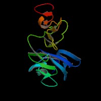
ModBase Predicted Comparative 3D Structure on P05107
Front
Top
Side
The pictures above may be empty if there is no ModBase structure for the protein. The ModBase structure frequently covers just a fragment of the protein. You may be asked to log onto ModBase the first time you click on the pictures. It is simplest after logging in to just click on the picture again to get to the specific info on that model.
Orthologous Genes in Other Species
Orthologies between human, mouse, and rat are computed by taking the best BLASTP hit, and filtering out non-syntenic hits. For more distant species reciprocal-best BLASTP hits are used. Note that the absence of an ortholog in the table below may reflect incomplete annotations in the other species rather than a true absence of the orthologous gene.
 US Server
 Sequence and Links to Tools and Databases
  Common Gene Haplotype Alleles