Description: Homo sapiens glutamate receptor, ionotropic, N-methyl D-aspartate 2B (GRIN2B), mRNA. RefSeq Summary (NM_000834): This gene encodes a member of the N-methyl-D-aspartate (NMDA) receptor family within the ionotropic glutamate receptor superfamily. The encoded protein is a subunit of the NMDA receptor ion channel which acts as an agonist binding site for glutamate. The NMDA receptors mediate a slow calcium-permeable component of excitatory synaptic transmission in the central nervous system. The NMDA receptors are heterotetramers of seven genetically encoded, differentially expressed subunits including NR1 (GRIN1), NR2 (GRIN2A, GRIN2B, GRIN2C, or GRIN2D) and NR3 (GRIN3A or GRIN3B). The early expression of this gene in development suggests a role in brain development, circuit formation, synaptic plasticity, and cellular migration and differentiation. Naturally occurring mutations within this gene are associated with neurodevelopmental disorders including autism spectrum disorder, attention deficit hyperactivity disorder, epilepsy, and schizophrenia. [provided by RefSeq, Aug 2017]. Transcript (Including UTRs) Position: hg19 chr12:13,714,410-14,133,022 Size: 418,613 Total Exon Count: 13 Strand: - Coding Region Position: hg19 chr12:13,715,717-14,019,142 Size: 303,426 Coding Exon Count: 12
ID:NMDE2_HUMAN DESCRIPTION: RecName: Full=Glutamate [NMDA] receptor subunit epsilon-2; AltName: Full=N-methyl D-aspartate receptor subtype 2B; Short=NMDAR2B; Short=NR2B; AltName: Full=N-methyl-D-aspartate receptor subunit 3; Short=NR3; Short=hNR3; Flags: Precursor; FUNCTION: NMDA receptor subtype of glutamate-gated ion channels with high calcium permeability and voltage-dependent sensitivity to magnesium. Mediated by glycine. In concert with DAPK1 at extrasynaptic sites, acts as a central mediator for stroke damage. Its phosphorylation at Ser-1303 by DAPK1 enhances synaptic NMDA receptor channel activity inducing injurious Ca2+ influx through them, resulting in an irreversible neuronal death (By similarity). SUBUNIT: Forms heteromeric channel of a zeta subunit (GRIN1), a epsilon subunit (GRIN2A, GRIN2B, GRIN2C or GRIN2D) and a third subunit (GRIN3A or GRIN3B). Found in a complex with GRIN1 and GRIN3B. Found in a complex with GRIN1, GRIN3A and PPP2CB. Interacts with PDZ domains of INADL and DLG4. Interacts with HIP1 and NETO1 (By similarity). Interacts with MAGI3. Interacts with DAPK1 (By similarity). SUBCELLULAR LOCATION: Cell membrane; Multi-pass membrane protein. Cell junction, synapse, postsynaptic cell membrane; Multi-pass membrane protein. TISSUE SPECIFICITY: Primarily found in the fronto-parieto-temporal cortex and hippocampus pyramidal cells, lower expression in the basal ganglia. PTM: Phosphorylation at Ser-1303 by DAPK1 enhances synaptic NMDA receptor channel activity (By similarity). DISEASE: Defects in GRIN2B are the cause of mental retardation autosomal dominant type 6 (MRD6) [MIM:613970]. Mental retardation is characterized by significantly below average general intellectual functioning associated with impairments in adaptative behavior and manifested during the developmental period. Note=Chromosomal aberrations involving GRIN2B have been found in patients with mental retardation. Translocations t(9;12)(p23;p13.1) and t(10;12)(q21.1;p13.1) with a common breakpoint in 12p13.1. SIMILARITY: Belongs to the glutamate-gated ion channel (TC 1.A.10.1) family. NR2B/GRIN2B subfamily.
alcoholism-related traits Wernicke C 2003, Polymorphisms in the N-methyl-D-aspartate receptor 1 and 2B subunits are associated with alcoholism-related traits., Biological psychiatry. 2003 Nov;54(9):922-8.
[PubMed 14573320]
These results suggest that variants in NMDAR genes are associated with alcoholism and related traits.
Alzheimer Disease S J Furney et al. Molecular psychiatry 2011, Genome-wide association with MRI atrophy measures as a quantitative trait locus for Alzheimer's disease., Molecular psychiatry.
[PubMed 21116278]
Amyotrophic Lateral Sclerosis Jennifer C Schymick et al. Lancet neurology 2007, Genome-wide genotyping in amyotrophic lateral sclerosis and neurologically normal controls: first stage analysis and public release of data., Lancet neurology.
[PubMed 17362836]
We generated publicly available genotype data for sporadic ALS patients and controls. No single locus was definitively associated with increased risk of developing disease, although potentially associated candidate SNPs were identified.
The RNAfold program from the Vienna RNA Package is used to perform the secondary structure predictions and folding calculations. The estimated folding energy is in kcal/mol. The more negative the energy, the more secondary structure the RNA is likely to have.
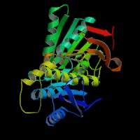
ModBase Predicted Comparative 3D Structure on Q13224
Front
Top
Side
The pictures above may be empty if there is no ModBase structure for the protein. The ModBase structure frequently covers just a fragment of the protein. You may be asked to log onto ModBase the first time you click on the pictures. It is simplest after logging in to just click on the picture again to get to the specific info on that model.
Orthologous Genes in Other Species
Orthologies between human, mouse, and rat are computed by taking the best BLASTP hit, and filtering out non-syntenic hits. For more distant species reciprocal-best BLASTP hits are used. Note that the absence of an ortholog in the table below may reflect incomplete annotations in the other species rather than a true absence of the orthologous gene.
 US Server