Description: Homo sapiens adenylate kinase 2 (AK2), nuclear gene encoding mitochondrial protein, transcript variant 1, mRNA. RefSeq Summary (NM_001625): Adenylate kinases are involved in regulating the adenine nucleotide composition within a cell by catalyzing the reversible transfer of phosphate groups among adenine nucleotides. Three isozymes of adenylate kinase, namely 1, 2, and 3, have been identified in vertebrates; this gene encodes isozyme 2. Expression of these isozymes is tissue-specific and developmentally regulated. Isozyme 2 is localized in the mitochondrial intermembrane space and may play a role in apoptosis. Mutations in this gene are the cause of reticular dysgenesis. Alternate splicing results in multiple transcript variants. Pseudogenes of this gene are found on chromosomes 1 and 2.[provided by RefSeq, Nov 2010]. Transcript (Including UTRs) Position: hg19 chr1:33,476,826-33,502,512 Size: 25,687 Total Exon Count: 6 Strand: - Coding Region Position: hg19 chr1:33,478,782-33,502,429 Size: 23,648 Coding Exon Count: 6
ID:KAD2_HUMAN DESCRIPTION: RecName: Full=Adenylate kinase 2, mitochondrial; Short=AK 2; EC=2.7.4.3; AltName: Full=ATP-AMP transphosphorylase 2; FUNCTION: Catalyzes the reversible transfer of the terminal phosphate group between ATP and AMP. This small ubiquitous enzyme involved in energy metabolism and nucleotide synthesis that is essential for maintenance and cell growth. Plays a key role in hematopoiesis. CATALYTIC ACTIVITY: ATP + AMP = 2 ADP. SUBUNIT: Monomer (By similarity). SUBCELLULAR LOCATION: Mitochondrion intermembrane space. TISSUE SPECIFICITY: Present in most tissues. Present at high level in heart, liver and kidney, and at low level in brain, skeletal muscle and skin. Present in thrombocytes but not in erythrocytes, which lack mitochondria. Present in all nucleated cell populations from blood, while AK1 is mostly absent. In spleen and lymph nodes, mononuclear cells lack AK1, whereas AK2 is readily detectable. These results indicate that leukocytes may be susceptible to defects caused by the lack of AK2, as they do not express AK1 in sufficient amounts to compensate for the AK2 functional deficits (at protein level). DISEASE: Defects in AK2 are the cause of reticular dysgenesis (RDYS) [MIM:267500]; also known as aleukocytosis. RDYS is the most severe form of inborn severe combined immunodeficiencies (SCID) and is characterized by absence of granulocytes and almost complete deficiency of lymphocytes in peripheral blood, hypoplasia of the thymus and secondary lymphoid organs, and lack of innate and adaptive humoral and cellular immune functions, leading to fatal septicemia within days after birth. In bone marrow of individuals with reticular dysgenesis, myeloid differentiation is blocked at the promyelocytic stage, whereas erythro- and megakaryocytic maturation is generally normal.In addition, affected newborns have bilateral sensorineural deafness. Defects may be due to its absence in leukocytes and inner ear, in which its absence can not be compensated by AK1. SIMILARITY: Belongs to the adenylate kinase family. AK2 subfamily.
The RNAfold program from the Vienna RNA Package is used to perform the secondary structure predictions and folding calculations. The estimated folding energy is in kcal/mol. The more negative the energy, the more secondary structure the RNA is likely to have.
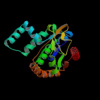
ModBase Predicted Comparative 3D Structure on P54819
Front
Top
Side
The pictures above may be empty if there is no ModBase structure for the protein. The ModBase structure frequently covers just a fragment of the protein. You may be asked to log onto ModBase the first time you click on the pictures. It is simplest after logging in to just click on the picture again to get to the specific info on that model.
Orthologous Genes in Other Species
Orthologies between human, mouse, and rat are computed by taking the best BLASTP hit, and filtering out non-syntenic hits. For more distant species reciprocal-best BLASTP hits are used. Note that the absence of an ortholog in the table below may reflect incomplete annotations in the other species rather than a true absence of the orthologous gene.
 US Server