Human Gene KIN (uc001ijt.3)
  Description: Homo sapiens KIN, antigenic determinant of recA protein homolog (mouse) (KIN), transcript variant 1, mRNA.
RefSeq Summary (NM_012311): The protein encoded by this gene is a nuclear protein that forms intranuclear foci during proliferation and is redistributed in the nucleoplasm during the cell cycle. Short-wave ultraviolet light provokes the relocalization of the protein, suggesting its participation in the cellular response to DNA damage. Originally selected based on protein-binding with RecA antibodies, the mouse protein presents a limited similarity with a functional domain of the bacterial RecA protein, a characteristic shared by this human ortholog. Alternative splicing of this gene results in multiple transcript variants. [provided by RefSeq, Jan 2012].
Transcript (Including UTRs)
   Position: hg19 chr10:7,792,925-7,829,990 Size: 37,066 Total Exon Count: 13 Strand: -
Coding Region
   Position: hg19 chr10:7,798,043-7,829,896 Size: 31,854 Coding Exon Count: 13 

Page IndexSequence and LinksUniProtKB CommentsPrimersGenetic AssociationsCTD
Gene AllelesRNA-Seq ExpressionMicroarray ExpressionRNA StructureProtein StructureOther Species
GO AnnotationsmRNA DescriptionsPathwaysOther NamesModel InformationMethods
Data last updated at UCSC: 2013-06-14

-  Sequence and Links to Tools and Databases
 
Genomic Sequence (chr10:7,792,925-7,829,990)mRNA (may differ from genome)Protein (393 aa)
Gene SorterGenome BrowserOther Species FASTAVisiGeneGene interactionsTable Schema
AlphaFoldBioGPSEnsemblEntrez GeneExonPrimerGeneCards
GeneNetworkH-INVHGNCHPRDLynxMGI
neXtProtOMIMPubMedReactomeTreefamUniProtKB
WikipediaBioGrid CRISPR DB

-  Comments and Description Text from UniProtKB
  ID: KIN17_HUMAN
DESCRIPTION: RecName: Full=DNA/RNA-binding protein KIN17; AltName: Full=Binding to curved DNA; AltName: Full=KIN, antigenic determinant of recA protein homolog;
FUNCTION: Involved in DNA replication and the cellular response to DNA damage. May participate in DNA replication factories and create a bridge between DNA replication and repair mediated by high molecular weight complexes. May play a role in illegitimate recombination and regulation of gene expression. May participate in mRNA processing. Binds, in vitro, to double-stranded DNA. Also shown to bind preferentially to curved DNA in vitro and in vivo (By similarity). Binds via its C-terminal domain to RNA in vitro.
SUBUNIT: Interacts with SV40 large T antigen. Associated with DNA polymerase alpha, RFC1 and cyclin A, in multiprotein DNA replication complexes. Also associates with replication origins at the G1/S phase boundary and throughout the S phase in vivo.
SUBCELLULAR LOCATION: Nucleus. Cytoplasm. Note=During S phase, strongly associated with the nuclear matrix, and to chromosomal DNA in the presence of DNA damage. Also shows cytoplasmic localization in elongated spermatids.
TISSUE SPECIFICITY: Ubiquitously expressed in all tissues examined, with highest levels in skeletal muscle, heart and testis. Differentially expressed in non-tumorigenic and tumorigenic cell lines. Highly expressed in proliferating epithelial keratinocyte cells in vitro (at protein level).
INDUCTION: By UVC irradiation in quiescent primary fibroblasts. By mitomycin C in human melanoma MeWO cells.
DOMAIN: The C-terminal domain (268-393) is organized into 2 subdomains that bear structural similarities to SH3-like domains. Both subdomains adopt a similar 5-stranded beta-barrel-like fold and are connected to each other by a short linker of 5 residues. The 5 beta-sheets are packed at approximately right angles against each other. A highly conserved groove formed at the interface between the 2 subdomains, comprised of Lys residues 302 and 391 and other positively charged residues, may possibly be the site of RNA-binding.
MISCELLANEOUS: Recognized by antibodies directed against the RecA protein (By similarity).
SIMILARITY: Belongs to the KIN17 family.
SIMILARITY: Contains 1 C2H2-type zinc finger.

-  Primer design for this transcript
 

Primer3Plus can design qPCR Primers that straddle exon-exon-junctions, which amplify only cDNA, not genomic DNA.
Click here to load the transcript sequence and exon structure into Primer3Plus

Exonprimer can design one pair of Sanger sequencing primers around every exon, located in non-genic sequence.
Click here to open Exonprimer with this transcript

To design primers for a non-coding sequence, zoom to a region of interest and select from the drop-down menu: View > In External Tools > Primer3


-  Genetic Association Studies of Complex Diseases and Disorders
  Genetic Association Database (archive): KIN
CDC HuGE Published Literature: KIN
Positive Disease Associations: monocyte chemoattractant protein 1 (66-77)
Related Studies:
  1. monocyte chemoattractant protein 1 (66-77)
    , , . [PubMed 0]

-  Comparative Toxicogenomics Database (CTD)
  The following chemicals interact with this gene           more ... click here to view the complete list

+  Common Gene Haplotype Alleles
  Press "+" in the title bar above to open this section.

-  RNA-Seq Expression Data from GTEx (53 Tissues, 570 Donors)
  Highest median expression: 3.24 RPKM in Brain - Cerebellar Hemisphere
Total median expression: 100.96 RPKM


View in GTEx track of Genome Browser    View at GTEx portal     View GTEx Body Map

+  Microarray Expression Data
  Press "+" in the title bar above to open this section.

-  mRNA Secondary Structure of 3' and 5' UTRs
 
RegionFold EnergyBasesEnergy/Base
Display As
5' UTR -39.1094-0.416 Picture PostScript Text
3' UTR -1434.355118-0.280 Picture PostScript Text

The RNAfold program from the Vienna RNA Package is used to perform the secondary structure predictions and folding calculations. The estimated folding energy is in kcal/mol. The more negative the energy, the more secondary structure the RNA is likely to have.

-  Protein Domain and Structure Information
  InterPro Domains: Graphical view of domain structure
IPR019447 - DNA/RNA-bd_Kin17_cons_domain
IPR015880 - Znf_C2H2-like

Pfam Domains:
PF10357 - Domain of Kin17 curved DNA-binding protein

Protein Data Bank (PDB) 3-D Structure
MuPIT help
2CKK - X-ray MuPIT 2V1N - NMR MuPIT


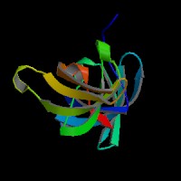
ModBase Predicted Comparative 3D Structure on O60870
FrontTopSide
The pictures above may be empty if there is no ModBase structure for the protein. The ModBase structure frequently covers just a fragment of the protein. You may be asked to log onto ModBase the first time you click on the pictures. It is simplest after logging in to just click on the picture again to get to the specific info on that model.

-  Orthologous Genes in Other Species
  Orthologies between human, mouse, and rat are computed by taking the best BLASTP hit, and filtering out non-syntenic hits. For more distant species reciprocal-best BLASTP hits are used. Note that the absence of an ortholog in the table below may reflect incomplete annotations in the other species rather than a true absence of the orthologous gene.
MouseRatZebrafishD. melanogasterC. elegansS. cerevisiae
No orthologNo orthologGenome BrowserGenome BrowserGenome BrowserGenome Browser
Gene Details  Gene DetailsGene DetailsGene Details
Gene Sorter  Gene SorterGene SorterGene Sorter
  EnsemblFlyBaseWormBaseSGD
  Protein SequenceProtein SequenceProtein SequenceProtein Sequence
  AlignmentAlignmentAlignmentAlignment

-  Gene Ontology (GO) Annotations with Structured Vocabulary
  Molecular Function:
GO:0003677 DNA binding
GO:0003690 double-stranded DNA binding
GO:0003723 RNA binding
GO:0005515 protein binding
GO:0046872 metal ion binding

Biological Process:
GO:0006260 DNA replication
GO:0006281 DNA repair
GO:0006310 DNA recombination
GO:0006397 mRNA processing
GO:0006974 cellular response to DNA damage stimulus
GO:0016032 viral process

Cellular Component:
GO:0005634 nucleus
GO:0005654 nucleoplasm
GO:0005737 cytoplasm
GO:0016363 nuclear matrix
GO:0032991 macromolecular complex
GO:0043231 intracellular membrane-bounded organelle


-  Descriptions from all associated GenBank mRNAs
  BC037913 - Homo sapiens cDNA clone IMAGE:5277557.
AK301789 - Homo sapiens cDNA FLJ58083 complete cds, highly similar to Homo sapiens KIN, antigenic determinant of recA protein homolog, mRNA.
JD480451 - Sequence 461475 from Patent EP1572962.
JD108079 - Sequence 89103 from Patent EP1572962.
BC017309 - Homo sapiens KIN, antigenic determinant of recA protein homolog (mouse), mRNA (cDNA clone MGC:29595 IMAGE:5089578), complete cds.
AJ005273 - Homo sapiens mRNA for Kin17 protein.
JD431720 - Sequence 412744 from Patent EP1572962.
KJ904679 - Synthetic construct Homo sapiens clone ccsbBroadEn_14073 KIN-like gene, encodes complete protein.
AK296637 - Homo sapiens cDNA FLJ51227 complete cds, highly similar to Homo sapiens KIN, antigenic determinant of recA protein homolog, mRNA.
AK309059 - Homo sapiens cDNA, FLJ99100.

-  Biochemical and Signaling Pathways
  Reactome (by CSHL, EBI, and GO)

Protein O60870 (Reactome details) participates in the following event(s):

R-HSA-8932275 METTL22 transfers 3xCH3 from 3xAdoMet to KIN
R-HSA-8876725 Protein methylation
R-HSA-597592 Post-translational protein modification
R-HSA-392499 Metabolism of proteins

-  Other Names for This Gene
  Alternate Gene Symbols: BTCD, KIN17, KIN17_HUMAN, NM_012311, NP_036443, O60870
UCSC ID: uc001ijt.3
RefSeq Accession: NM_012311
Protein: O60870 (aka KIN17_HUMAN)
CCDS: CCDS7080.1

-  Gene Model Information
 
category: coding nonsense-mediated-decay: no RNA accession: NM_012311.3
exon count: 13CDS single in 3' UTR: no RNA size: 6401
ORF size: 1182CDS single in intron: no Alignment % ID: 100.00
txCdsPredict score: 2564.00frame shift in genome: no % Coverage: 99.89
has start codon: yes stop codon in genome: no # of Alignments: 1
has end codon: yes retained intron: yes # AT/AC introns 0
selenocysteine: no end bleed into intron: 0# strange splices: 0
Click here for a detailed description of the fields of the table above.

-  Methods, Credits, and Use Restrictions
  Click here for details on how this gene model was made and data restrictions if any.