Description: Homo sapiens protocadherin 12 (PCDH12), mRNA. RefSeq Summary (NM_016580): This gene belongs to the protocadherin gene family, a subfamily of the cadherin superfamily. The encoded protein consists of an extracellular domain containing 6 cadherin repeats, a transmembrane domain and a cytoplasmic tail that differs from those of the classical cadherins. The gene localizes to the region on chromosome 5 where the protocadherin gene clusters reside. The exon organization of this transcript is similar to that of the gene cluster transcripts, notably the first large exon, but no significant sequence homology exists. The function of this cellular adhesion protein is undetermined but mouse protocadherin 12 does not bind catenins and appears to have no affect on cell migration or growth. [provided by RefSeq, Jul 2008]. Transcript (Including UTRs) Position: hg19 chr5:141,324,530-141,338,627 Size: 14,098 Total Exon Count: 4 Strand: - Coding Region Position: hg19 chr5:141,324,946-141,337,416 Size: 12,471 Coding Exon Count: 4
ID:PCD12_HUMAN DESCRIPTION: RecName: Full=Protocadherin-12; AltName: Full=Vascular cadherin-2; AltName: Full=Vascular endothelial cadherin-2; Short=VE-cad-2; Short=VE-cadherin-2; Flags: Precursor; FUNCTION: Cellular adhesion molecule that may play an important role in cell-cell interactions at interendothelial junctions. Promotes homotypic calcium-dependent aggregation and adhesion and clusters at intercellular junctions. Unable to bind to catenins, weakly associates with the cytoskeleton (By similarity). SUBCELLULAR LOCATION: Cell membrane; Single-pass type I membrane protein. Cell junction. TISSUE SPECIFICITY: Expressed in highly vascularized tissues including the heart and placenta, but most tissues contain a low level of expression. Prominent expression in the spleen. SIMILARITY: Contains 6 cadherin domains.
The RNAfold program from the Vienna RNA Package is used to perform the secondary structure predictions and folding calculations. The estimated folding energy is in kcal/mol. The more negative the energy, the more secondary structure the RNA is likely to have.
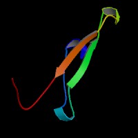
ModBase Predicted Comparative 3D Structure on Q9NPG4
Front
Top
Side
The pictures above may be empty if there is no ModBase structure for the protein. The ModBase structure frequently covers just a fragment of the protein. You may be asked to log onto ModBase the first time you click on the pictures. It is simplest after logging in to just click on the picture again to get to the specific info on that model.
Orthologous Genes in Other Species
Orthologies between human, mouse, and rat are computed by taking the best BLASTP hit, and filtering out non-syntenic hits. For more distant species reciprocal-best BLASTP hits are used. Note that the absence of an ortholog in the table below may reflect incomplete annotations in the other species rather than a true absence of the orthologous gene.
 US Server