Description: Homo sapiens prion protein (PRNP), transcript variant 5, mRNA. RefSeq Summary (NM_001080123): The protein encoded by this gene is a membrane glycosylphosphatidylinositol-anchored glycoprotein that tends to aggregate into rod-like structures. The encoded protein contains a highly unstable region of five tandem octapeptide repeats. This gene is found on chromosome 20, approximately 20 kbp upstream of a gene which encodes a biochemically and structurally similar protein to the one encoded by this gene. Mutations in the repeat region as well as elsewhere in this gene have been associated with Creutzfeldt-Jakob disease, fatal familial insomnia, Gerstmann-Straussler disease, Huntington disease-like 1, and kuru. An overlapping open reading frame has been found for this gene that encodes a smaller, structurally unrelated protein, AltPrp. Alternative splicing results in multiple transcript variants. [provided by RefSeq, Nov 2014]. Transcript (Including UTRs) Position: hg19 chr20:4,667,157-4,682,234 Size: 15,078 Total Exon Count: 2 Strand: + Coding Region Position: hg19 chr20:4,679,867-4,680,628 Size: 762 Coding Exon Count: 1
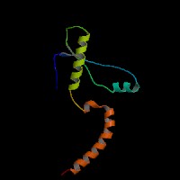
ID:PRIO_HUMAN DESCRIPTION: RecName: Full=Major prion protein; Short=PrP; AltName: Full=ASCR; AltName: Full=PrP27-30; AltName: Full=PrP33-35C; AltName: CD_antigen=CD230; Flags: Precursor; FUNCTION: May play a role in neuronal development and synaptic plasticity. May be required for neuronal myelin sheath maintenance. May play a role in iron uptake and iron homeostasis. Soluble oligomers are toxic to cultured neuroblastoma cells and induce apoptosis (in vitro). Association with GPC1 (via its heparan sulfate chains) targets PRNP to lipid rafts. Also provides Cu(2+) or ZN(2+) for the ascorbate-mediated GPC1 deaminase degradation of its heparan sulfate side chains (By similarity). SUBUNIT: Monomer and homodimer. Has a tendency to aggregate into amyloid fibrils containing a cross-beta spine, formed by a steric zipper of superposed beta-strands. Soluble oligomers may represent an intermediate stage on the path to fibril formation. Copper binding may promote oligomerization. Interacts with GRB2, APP, ERI3/PRNPIP and SYN1. Mislocalized cytosolically exposed PrP interacts with MGRN1; this interaction alters MGRN1 subcellular location and causes lysosomal enlargement (By similarity). Interacts with KIAA1191. INTERACTION: Self; NbExp=10; IntAct=EBI-977302, EBI-977302; Q9BSJ6:FAM64A; NbExp=5; IntAct=EBI-977302, EBI-2568609; P49639:HOXA1; NbExp=4; IntAct=EBI-977302, EBI-740785; P29372:MPG; NbExp=4; IntAct=EBI-977302, EBI-1043398; Q9H4B4:PLK3; NbExp=4; IntAct=EBI-977302, EBI-751877; SUBCELLULAR LOCATION: Cell membrane; Lipid-anchor, GPI-anchor. Golgi apparatus. Note=Targeted to lipid rafts via association with the heparan sulfate chains of GPC1. Colocates, in the presence of CU(2+), to vesicles in para- and perinuclear regions, where both proteins undergo internalization. Heparin displaces PRNP from lipid rafts and promotes endocytosis. SUBCELLULAR LOCATION: Isoform 2: Cytoplasm. Nucleus. Note=Accumulates outside the secretory route in the cytoplasm, from where it relocates to the nucleus. DOMAIN: The normal, monomeric form, PRPN(C), has a mainly alpha- helical structure. Misfolding of this form produces a disease- associated, protease-resistant form, PRPN (Sc), accompanied by a large increase of the beta-sheet content and formation of amyloid fibrils. These fibrils consist of a cross-beta spine, formed by a steric zipper of superposed beta-strands. Disease mutations may favor intermolecular contacts via short beta strands, and may thereby trigger oligomerization. In addition, the heparan-sulfate proteoglycan, GPC1, promotes the association of PRPN (C) to lipid rafts and appears to facilitate the conversion to PRPN (Sc). DOMAIN: Contains an N-terminal region composed of octamer repeats. At low copper concentrations, the sidechains of His residues from three or four repeats contribute to the binding of a single copper ion. Alternatively, a copper ion can be bound by interaction with the sidechain and backbone amide nitrogen of a single His residue. The observed copper binding stoichiometry suggests that two repeat regions cooperate to stabilize the binding of a single copper ion. At higher copper concentrations, each octamer can bind one copper ion by interactions with the His sidechain and Gly backbone atoms. A mixture of binding types may occur, especially in the case of octamer repeat expansion. Copper binding may stabilize the conformation of this region and may promote oligomerization. PTM: The glycosylation pattern (the amount of mono-, di- and non- glycosylated forms or glycoforms) seems to differ in normal and CJD prion. PTM: Isoform 2 is sumoylated with SUMO1 (By similarity). POLYMORPHISM: The five tandem octapeptide repeats region is highly unstable. Insertions or deletions of octapeptide repeat units are associated to prion disease. DISEASE: Note=PrP is found in high quantity in the brain of humans and animals infected with neurodegenerative diseases known as transmissible spongiform encephalopathies or prion diseases, like: Creutzfeldt-Jakob disease (CJD), fatal familial insomnia (FFI), Gerstmann-Straussler disease (GSD), Huntington disease-like type 1 (HDL1) and kuru in humans; scrapie in sheep and goat; bovine spongiform encephalopathy (BSE) in cattle; transmissible mink encephalopathy (TME); chronic wasting disease (CWD) of mule deer and elk; feline spongiform encephalopathy (FSE) in cats and exotic ungulate encephalopathy (EUE) in nyala and greater kudu. The prion diseases illustrate three manifestations of CNS degeneration: (1) infectious (2) sporadic and (3) dominantly inherited forms. TME, CWD, BSE, FSE, EUE are all thought to occur after consumption of prion-infected foodstuffs. DISEASE: Defects in PRNP are the cause of Creutzfeldt-Jakob disease (CJD) [MIM:123400]. CJD occurs primarily as a sporadic disorder (1 per million), while 10-15% are familial. Accidental transmission of CJD to humans appears to be iatrogenic (contaminated human growth hormone (HGH), corneal transplantation, electroencephalographic electrode implantation, etc.). Epidemiologic studies have failed to implicate the ingestion of infected annimal meat in the pathogenesis of CJD in human. The triad of microscopic features that characterize the prion diseases consists of (1) spongiform degeneration of neurons, (2) severe astrocytic gliosis that often appears to be out of proportion to the degree of nerve cell loss, and (3) amyloid plaque formation. CJD is characterized by progressive dementia and myoclonic seizures, affecting adults in mid-life. Some patients present sleep disorders, abnormalities of high cortical function, cerebellar and corticospinal disturbances. The disease ends in death after a 3-12 months illness. DISEASE: Defects in PRNP are the cause of fatal familial insomnia (FFI) [MIM:600072]. FFI is an autosomal dominant disorder and is characterized by neuronal degeneration limited to selected thalamic nuclei and progressive insomnia. DISEASE: Defects in PRNP are the cause of Gerstmann-Straussler disease (GSD) [MIM:137440]. GSD is a heterogeneous disorder and was defined as a spinocerebellar ataxia with dementia and plaquelike deposits. GSD incidence is less than 2 per 100 million live births. DISEASE: Defects in PRNP are the cause of Huntington disease-like type 1 (HDL1) [MIM:603218]. HDL1 is an autosomal dominant, early onset neurodegenerative disorder with prominent psychiatric features. DISEASE: Defects in PRNP are the cause of kuru (KURU) [MIM:245300]. Kuru is transmitted during ritualistic cannibalism, among natives of the New Guinea highlands. Patients exhibit various movement disorders like cerebellar abnormalities, rigidity of the limbs, and clonus. Emotional lability is present, and dementia is conspicuously absent. Death usually occurs from 3 to 12 month after onset. DISEASE: Defects in PRNP are the cause of spongiform encephalopathy with neuropsychiatric features (SENF) [MIM:606688]; an autosomal dominant presenile dementia with a rapidly progressive and protracted clinical course. The dementia was characterized clinically by frontotemporal features, including early personality changes. Some patients had memory loss, several showed aggressiveness, hyperorality and verbal stereotypy, others had parkinsonian symptoms. MISCELLANEOUS: This protein is produced by a bicistronic gene which also produces the The alternative prion protein/AltPrP from an overlapping reading frame. MISCELLANEOUS: The alternative prion protein/AltPrP (AC F7VJQ1) and PRNP have no apparent direct functional relation since a mutation that removes the start codon of the AltPrP has no apparent effect on the biology of PRNP. In mouse and hamster, the alternative initiation AUG codon is absent and is replaced by a GUG codon. SIMILARITY: Belongs to the prion family. WEB RESOURCE: Name=The Official Mad Cow Disease Home Page; URL="http://www.mad-cow.org/"; WEB RESOURCE: Name=GeneReviews; URL="http://www.ncbi.nlm.nih.gov/sites/GeneTests/lab/gene/PRNP"; WEB RESOURCE: Name=Wikipedia; Note=PRNP entry; URL="http://en.wikipedia.org/wiki/PRNP";
Alzheimer's Disease Croes, E. A. et al. 2004, Octapeptide repeat insertions in the prion protein gene and early onset dementia., Journal of neurology, neurosurgery, and psychiatry. 2004 Aug;75(8):1166-70.
[PubMed 15258222]
Our findings show significant inverse associations of the length of the PRNP octapeptide repeat with age at disease onset and disease duration in the spongiform encephalopathies.
Alzheimer's Disease Del Bo, R. et al. 2005, Is M129V of PRNP gene associated with Alzheimer's disease? A case-control study and a meta-analysis., Neurobiology of aging. 2005.
[PubMed 16099550]
cerebral aging Berr 2003, Polymorphism of the codon 129 of the prion protein (PrP) gene and neuropathology of cerebral ageing., Acta neuropathologica. 2003 Jul;106(1):71-4.
[PubMed 12679875]
Val allele carriers also had more focal and diffuse Abeta deposits. This association was most significant in the highest Braak's stages for neurofibrillary tangles (>/=III). In this group, cases with at least one Val allele had nearly twice as many Abeta-associated lesions. The most affected areas were the entorhinal cortex, TF-TH and the superior temporal cortex, where odds ratios for focal Abeta deposits ranged from 3.5 to 4.6.
The RNAfold program from the Vienna RNA Package is used to perform the secondary structure predictions and folding calculations. The estimated folding energy is in kcal/mol. The more negative the energy, the more secondary structure the RNA is likely to have.
ModBase Predicted Comparative 3D Structure on P04156
Front
Top
Side
The pictures above may be empty if there is no ModBase structure for the protein. The ModBase structure frequently covers just a fragment of the protein. You may be asked to log onto ModBase the first time you click on the pictures. It is simplest after logging in to just click on the picture again to get to the specific info on that model.
Orthologous Genes in Other Species
Orthologies between human, mouse, and rat are computed by taking the best BLASTP hit, and filtering out non-syntenic hits. For more distant species reciprocal-best BLASTP hits are used. Note that the absence of an ortholog in the table below may reflect incomplete annotations in the other species rather than a true absence of the orthologous gene.
Biological Process: GO:0001933 negative regulation of protein phosphorylation GO:0006878 cellular copper ion homeostasis GO:0006979 response to oxidative stress GO:0007049 cell cycle GO:0007050 cell cycle arrest GO:0007611 learning or memory GO:0007616 long-term memory GO:0010942 positive regulation of cell death GO:0010955 negative regulation of protein processing GO:0031648 protein destabilization GO:0032147 activation of protein kinase activity GO:0032689 negative regulation of interferon-gamma production GO:0032700 negative regulation of interleukin-17 production GO:0032703 negative regulation of interleukin-2 production GO:0032880 regulation of protein localization GO:0035584 calcium-mediated signaling using intracellular calcium source GO:0035690 cellular response to drug GO:0043066 negative regulation of apoptotic process GO:0043086 negative regulation of catalytic activity GO:0043433 negative regulation of sequence-specific DNA binding transcription factor activity GO:0043525 positive regulation of neuron apoptotic process GO:0046007 negative regulation of activated T cell proliferation GO:0046686 response to cadmium ion GO:0046688 response to copper ion GO:0050730 regulation of peptidyl-tyrosine phosphorylation GO:0050731 positive regulation of peptidyl-tyrosine phosphorylation GO:0050860 negative regulation of T cell receptor signaling pathway GO:0051260 protein homooligomerization GO:0061098 positive regulation of protein tyrosine kinase activity GO:0070885 negative regulation of calcineurin-NFAT signaling cascade GO:0071280 cellular response to copper ion GO:0090314 positive regulation of protein targeting to membrane GO:0090647 modulation of age-related behavioral decline GO:0097062 dendritic spine maintenance GO:1900272 negative regulation of long-term synaptic potentiation GO:1900449 regulation of glutamate receptor signaling pathway GO:1901216 positive regulation of neuron death GO:1901379 regulation of potassium ion transmembrane transport GO:1902430 negative regulation of beta-amyloid formation GO:1902938 regulation of intracellular calcium activated chloride channel activity GO:1902951 negative regulation of dendritic spine maintenance GO:1902992 negative regulation of amyloid precursor protein catabolic process GO:1903078 positive regulation of protein localization to plasma membrane GO:1904645 response to beta-amyloid GO:1904646 cellular response to beta-amyloid GO:1905664 regulation of calcium ion import across plasma membrane GO:1990535 neuron projection maintenance
 US Server