Description: Homo sapiens interleukin 1 receptor-like 2 (IL1RL2), mRNA. RefSeq Summary (NM_003854): The protein encoded by this gene is a member of the interleukin 1 receptor family. An experiment with transient gene expression demonstrated that this receptor was incapable of binding to interleukin 1 alpha and interleukin 1 beta with high affinity. This gene and four other interleukin 1 receptor family genes, including interleukin 1 receptor, type I (IL1R1), interleukin 1 receptor, type II (IL1R2), interleukin 1 receptor-like 1 (IL1RL1), and interleukin 18 receptor 1 (IL18R1), form a cytokine receptor gene cluster in a region mapped to chromosome 2q12. [provided by RefSeq, Jul 2008]. Transcript (Including UTRs) Position: hg19 chr2:102,803,433-102,855,811 Size: 52,379 Total Exon Count: 12 Strand: + Coding Region Position: hg19 chr2:102,804,328-102,855,701 Size: 51,374 Coding Exon Count: 11
ID:ILRL2_HUMAN DESCRIPTION: RecName: Full=Interleukin-1 receptor-like 2; AltName: Full=Interleukin-1 receptor-related protein 2; Short=IL-1Rrp2; Short=IL1R-rp2; Flags: Precursor; FUNCTION: Receptor for interleukin 1 family member 9 (IL1F9). Binding to the agonist leads to the activation of NF-kappa-B. SUBUNIT: Does not bind IL1A/interleukin-1 alpha or IL1B/interleukin-1 beta. SUBCELLULAR LOCATION: Membrane; Single-pass type I membrane protein. SIMILARITY: Belongs to the interleukin-1 receptor family. SIMILARITY: Contains 3 Ig-like C2-type (immunoglobulin-like) domains. SIMILARITY: Contains 1 TIR domain. SEQUENCE CAUTION: Sequence=AAB53237.1; Type=Frameshift; Positions=556; WEB RESOURCE: Name=SeattleSNPs; URL="http://pga.gs.washington.edu/data/il1rl2/";
Parkinson Disease Hon-Chung Fung et al. Lancet neurology 2006, Genome-wide genotyping in Parkinson's disease and neurologically normal controls: first stage analysis and public release of data., Lancet neurology.
[PubMed 17052657]
We generated publicly available genotype data for Parkinsons disease patients and controls so that these data can be mined and augmented by other researchers to identify common genetic variability that results in minor and moderate risk for disease.
The RNAfold program from the Vienna RNA Package is used to perform the secondary structure predictions and folding calculations. The estimated folding energy is in kcal/mol. The more negative the energy, the more secondary structure the RNA is likely to have.
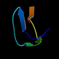
ModBase Predicted Comparative 3D Structure on Q9HB29
Front
Top
Side
The pictures above may be empty if there is no ModBase structure for the protein. The ModBase structure frequently covers just a fragment of the protein. You may be asked to log onto ModBase the first time you click on the pictures. It is simplest after logging in to just click on the picture again to get to the specific info on that model.
Orthologous Genes in Other Species
Orthologies between human, mouse, and rat are computed by taking the best BLASTP hit, and filtering out non-syntenic hits. For more distant species reciprocal-best BLASTP hits are used. Note that the absence of an ortholog in the table below may reflect incomplete annotations in the other species rather than a true absence of the orthologous gene.
 US Server